The eyes are the first thing many of us notice when we meet someone new. We think of them as windows to the soul and spend plenty of time staring into the eyes of people we love. But do you know how eyes work? Do you know about common vision problems that can affect you and your loved ones?
Our eyes are hard at work all the time. They give us valuable information about the world around us, creating images like a highly advanced camera.
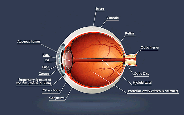
The human eye consists of many different and intricate parts, all working together to provide you with sight. The eye works much like a camera. It is a light-gathering device that constructs images of the world around you as rays reflect off of objects in the environment and travel through your cornea.

The major parts of the eye include:
Aqueous Humor
The aqueous humor is a clear fluid that fills the space between the lens and cornea (front of the eyeball). The fluid bathes and nourishes the lens and maintains pressure within the eye.
Cornea
At the very front of your eye is a transparent, dome-shaped lens called the cornea. It covers and protects the iris and plays a vital role in providing clear vision. As light rays reflect off of objects in the environment, they enter the eyes through the cornea. The cornea then refracts (bends) the rays as they pass through the pupil.
Conjunctiva
The conjunctiva is the thin transparent layer of tissue that lines the inner surface of the eyelid that covers the white part of the eye. Its function is to keep the eye moist and lubricated.
Fovea
The center of the macula, where your vision is sharpest.
Macula
The macula is the small, sensitive area of the retina needed for central vision. It contains the fovea.
Iris
The iris is the colored part of your eye that divides the front from the back. This iris is embedded with small muscles that dilate and constrict the pupil size based on the amount of available light. In bright light, the iris's sphincter muscles tighten and cause the pupil to contract. In dim light, dilator muscles will increase the size of the pupil.
Lens
The crystalline lens is located just behind the iris. As light comes through the pupil, the lens focuses this light onto the retina. As it focuses, the lens changes shape to adjust for the closeness or distance of the objects through a process called accommodation. As we grow older, the lens gradually hardens and the ability to accommodate slowly diminishes.
Optic Nerve
The optic nerve is a bundle of more than 1 million nerve fibers that carry visual messages from the retina to the brain.
Pupil
The pupil is the round opening in the center of your eye. Its size determines the amount of light that will enter the eye. The pupil works with the dilator and the sphincter muscles of the iris to change size to let more or less light in.
Retina
The retina is a sensory tissue that lines the back of the eye. It is filled with two types of photoreceptors, rods, and cones, which work to create central and peripheral vision. The retina captures light rays and converts them to electrical impulses. These impulses travel along the optic nerve to the brain, where they become the images that we see around us.
Sclera
The white outer coating of the eye.
Vitreous Humor
The vitreous humour is a clear, colorless fluid or gel that fills the space between the lens and the retina of your eye.


